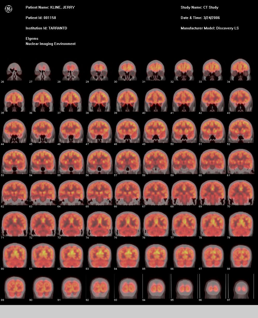
27 March 2006: Here are images from my PET scan performed on Friday, 17 March 2006. This first one is the Coronal (back view) Fused.
The lighter the color, the heavier the uptake (glucose consumption).
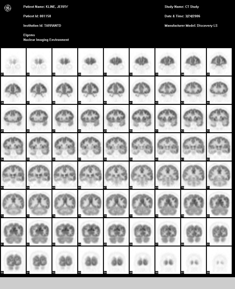
Raw PET frontal images. Fused coloring is inverted from PET images. Darker PET regions equate to higher uptake activity, and lighter areas indicate less uptake.
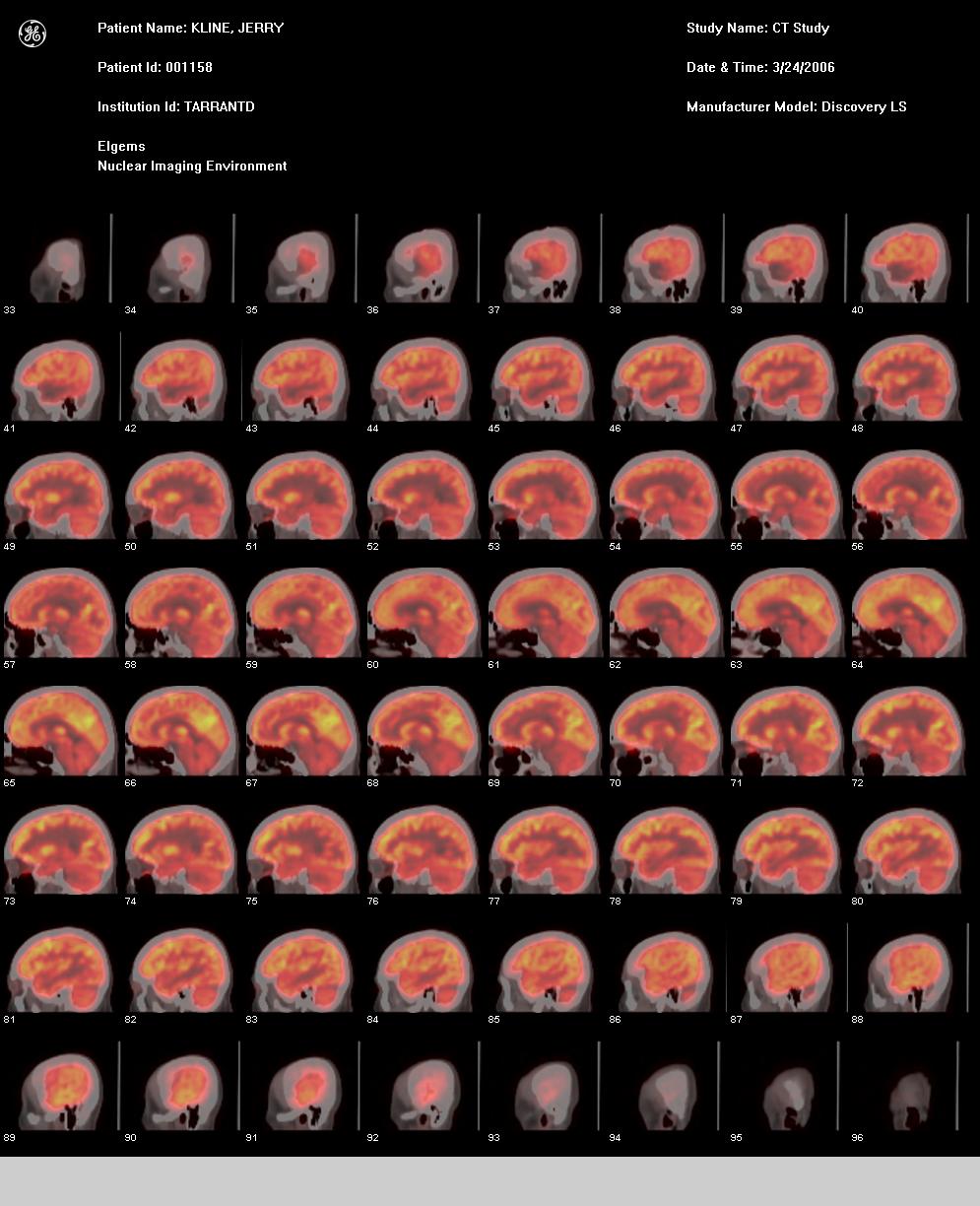
Sagittal (side view) Fused images. Again, lighter colored areas show which parts of my brain are in use.
Dr. Stark-Vance says that you can even tell by looking at these pics what I was doing at the time - moving my right foot, looking at something, etc.
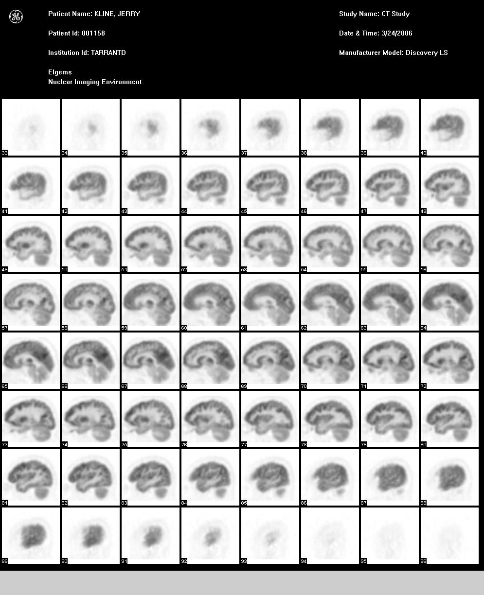
Sagittal PET images. So the reason that the patient is to chill for 90 minutes prior to the scan is to minimize the brain activity and keep as much of the injected FDG available to find and monitor any abnormal activity, such as the higher sugar consumption of a tumor.
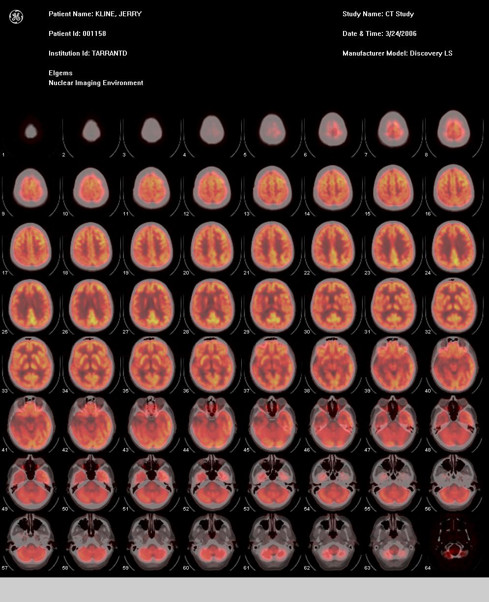
Transaxial (top view) Fused images. Key elements to look for are color symmetry. Notice the dark areas representing my eyes.
I had my eyes closed, so minimal glucose consumption was occurring.

Transaxial PET images. I now have proof that there is indeed something (good) in my head, contrary to popular belief!